
Finding a breast lump can be unsettling. Even a small change may bring an immediate rush of worry, especially because breast lumps are so closely associated with cancer in public conversation. Yet many lumps are caused by noncancerous conditions, including cysts, fibroadenomas, hormonal changes, and infections. The important point is not to guess which type it might be based on touch alone.
Understanding breast lump types and diagnosis can make the evaluation process feel less mysterious. A lump’s texture, location, tenderness, and relationship to the menstrual cycle may provide useful clues, but these features cannot confirm the cause. Any new or unexplained breast lump should be assessed by a qualified healthcare professional.
What counts as a breast lump
A breast lump is an area that feels different from the tissue around it. It may be round or irregular, soft or firm, painful or completely painless. Some lumps move slightly beneath the fingers, while others seem fixed in place. A person may also notice general thickening rather than one clearly defined mass.
Breast tissue naturally has texture. Hormonal changes, age, pregnancy, breastfeeding, and menopause can all affect how the breasts feel. One breast may also be slightly different from the other without anything being wrong. The concern usually begins when there is a new, persistent, or noticeably changing area.
Most breast changes are not cancer, but the National Cancer Institute and the NHS advise having unusual changes checked rather than attempting to diagnose them at home.
Fibrocystic breast changes
Fibrocystic breast changes can cause tenderness, swelling, and areas of lumpiness, often in both breasts. The texture may become more noticeable before a period and improve once menstruation begins. Rather than feeling like one separate lump, the breast may feel generally rope-like, nodular, or uneven.
These changes are benign and are especially common during the reproductive years. They are influenced by hormonal fluctuations and often become less noticeable after menopause. Ordinary fibrocystic changes do not increase breast cancer risk, although any distinct new lump within generally lumpy tissue still needs evaluation.
Breast cysts
A breast cyst is a fluid-filled sac within the breast. Cysts may feel smooth, round, or oval, and some are tender when pressed. Their size and sensitivity can change during the menstrual cycle. They are particularly common before menopause, though they can occur at other ages as well.
Ultrasound is often used to determine whether a lump is filled with fluid or is solid. A simple cyst may require no treatment if it is not causing discomfort. When a cyst is painful or unusually large, a clinician may drain it using a fine needle. The fluid removal can relieve pressure and may cause the lump to collapse.
Fibroadenomas
A fibroadenoma is a solid, noncancerous growth made from glandular and connective breast tissue. It often feels smooth, firm, rubbery, and well defined. Many fibroadenomas move easily beneath the skin when touched, which is why they are sometimes described as mobile lumps.
They are most common in younger women, particularly between the teenage years and the mid-thirties, but they can appear at any age. Some remain the same size, while others grow during pregnancy or periods of hormonal change. Imaging may strongly suggest a fibroadenoma, but follow-up or biopsy may be recommended when its appearance, size, or growth pattern needs clarification.
Infections and breast abscesses
Mastitis is inflammation of the breast that is often associated with breastfeeding, although it can occur in people who are not breastfeeding. The affected area may become painful, swollen, hot, or red. Fever, chills, tiredness, and a generally unwell feeling may develop as well.
If infected fluid collects within the tissue, a breast abscess can form. This may feel like a painful lump and usually requires prompt medical treatment. Antibiotics may be needed, and an abscess sometimes has to be drained. A hot, rapidly swelling breast accompanied by fever should not be treated as an ordinary hormonal lump.
Intraductal papillomas
An intraductal papilloma is a small, usually benign growth inside a milk duct. It often develops near the nipple and may be too small to feel clearly. One of its more noticeable signs is clear or bloody nipple discharge, sometimes from only one breast.
Although an intraductal papilloma is not necessarily cancerous, nipple discharge that occurs without squeezing, especially when it contains blood, should be investigated. Ultrasound, mammography, or other tests may be used to examine the duct and surrounding tissue.
Fat necrosis and other benign lumps
Fat necrosis can occur when fatty breast tissue is damaged by an injury, surgery, radiation treatment, or another form of trauma. The damaged area may harden into a lump. Sometimes the original injury was minor or happened long enough ago that the person no longer remembers it.
Other noncancerous lumps include lipomas, which are made of fatty tissue, and adenosis, in which the breast lobules become enlarged. These conditions can sometimes resemble more concerning changes during a physical examination or on imaging. That overlap is one reason diagnosis may require more than simply feeling the area. The NCI describes several benign breast conditions that vary in their symptoms and their relationship to future cancer risk.
Breast cancer and suspicious changes
A cancerous breast lump is often described as hard or irregular, but breast cancer does not always follow a predictable pattern. A malignant lump may be painless, tender, smooth, or difficult to distinguish from benign tissue. Pain or mobility cannot reliably rule cancer in or out.
Warning signs can include a new lump in the breast or armpit, skin dimpling, unexplained redness, a change in breast shape, nipple inversion, persistent nipple rash, or discharge that is not breast milk. Inflammatory breast cancer may cause rapid swelling, warmth, redness, or skin that resembles orange peel, sometimes without a distinct lump.
How a breast lump is examined
Diagnosis usually begins with a discussion of when the lump was noticed, whether it has changed, and whether it varies with the menstrual cycle. The clinician may ask about pain, nipple discharge, pregnancy, breastfeeding, medicines, previous breast problems, and family history.
A physical examination follows. The clinician checks the lump’s location, size, texture, and mobility while also examining the skin, nipples, other breast, and lymph nodes around the armpit and collarbone. These observations help guide the next step, but an examination alone may not provide a definite answer.
Mammography and breast ultrasound
The first imaging test often depends on the person’s age, symptoms, pregnancy status, and medical history. Ultrasound uses sound waves to show whether a lump is solid or fluid-filled. It is commonly the first imaging method for younger women, pregnant or breastfeeding patients, and people whose breast tissue is relatively dense.
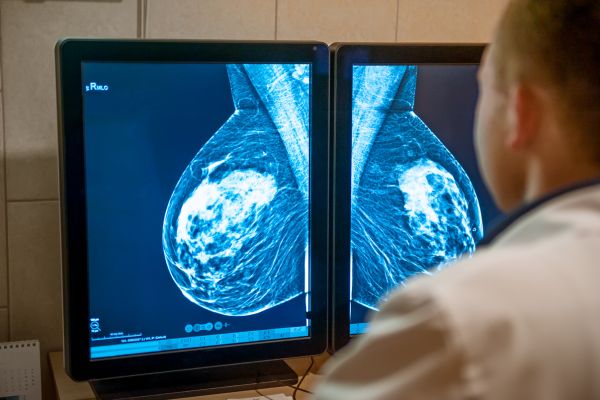
Diagnostic mammography creates detailed X-ray images of the breast and is commonly used when evaluating a new lump in women aged 40 or older. Women between 30 and 39 may have ultrasound, mammography, or both, depending on the clinical situation. Imaging recommendations are individualized, so these age ranges should not be treated as rigid rules.
When a biopsy is needed
If imaging shows an uncertain or suspicious area, a biopsy may be recommended. During a core needle biopsy, a hollow needle removes small samples of tissue, usually with guidance from ultrasound or mammography. A pathologist then examines the samples under a microscope.
A biopsy does not automatically mean cancer is expected. It is a method of obtaining a definite tissue diagnosis when examination and imaging cannot provide enough certainty. Some lumps are monitored with repeat imaging instead, particularly when they have features that appear probably benign.
Why every new lump deserves attention
Most breast lumps turn out to be benign, particularly in younger women. Still, appearance and texture are not reliable enough for self-diagnosis. A harmless cyst can feel firm, while a cancerous lump may not match the classic descriptions found online.
Breast lump types and diagnosis involve bringing together the clinical examination, imaging findings, and, when necessary, biopsy results. Seeking an assessment does not mean assuming the worst. It means replacing uncertainty with evidence.
A new lump, persistent thickening, nipple change, unusual discharge, skin dimpling, or swelling in the armpit should be checked without unnecessary delay. Being familiar with the normal look and feel of your breasts can help you notice changes earlier, but professional evaluation is what determines their meaning. Most findings bring reassurance; those that require treatment are better addressed when they are recognized promptly.